


Osteochondritis Dissecans
Osteochondritis dissecans is a joint condition in which a piece of cartilage, along with a thin layer of the bone separates from the end of the bone because of inadequate blood supply. The separated fragments are sometimes called “joint mice”. These fragments may be localized, or may detach and fall into the joint space causing pain and joint instability.
Anatomy
The knee, mostly the femoral condyles are most commonly affected. The two femoral condyles make up for the rounded end of femur (thigh bone). Each knee has two femoral condyles, the medial femoral condyle on the inside of the knee and the lateral femoral condyle on the outside of the knee. Osteochondritis dissecans occurs within the lateral aspect of the medial femoral condyle. The condition can also occur in other joints, including your elbows, ankles, shoulders and hips.
Incidence
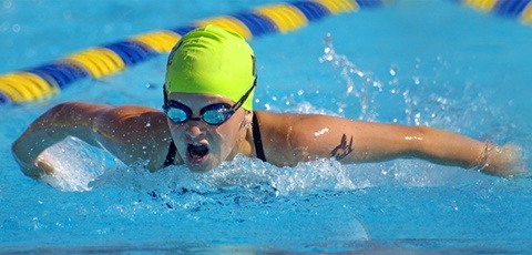
Osteochondritis dissecans is more common among boys and young men between 10 and 20 years who actively take part in sports Athletes participating in sports such as gymnastics and baseball may develop osteochondritis dissecans.
Causes
Exact cause for osteochondritis dissecans remains unknown and certain factors such as trauma, fractures, sprains, or injury to the joint are considered to increase the risk of developing the condition. Osteochondritis dissecans may be caused by restricted blood supply to the end of the affected bone that usually occurs in conjunction with repetitive trauma. Following the injury or trauma, the bones in the area may be deprived of blood flow leading to necrosis and finally the bone fragment may break off. This may initiate the healing process however by this time, articular cartilage will be compressed, flattened, and a subchondral cyst will be developed. All these changes in addition to increased joint pressure cause failure of healing of the joint.
The appearance of osteochondritis dissecans in several family members may indicate that the condition is inherited.
Symptoms
Patients with osteochondritis dissecans usually have joint pain, swelling, stiffness, decreased range of motion, and joint popping or locking. Pain usually increases after activity.
Diagnosis
Your doctor will probably order an X-ray of both the right and left knee to see the abnormality in the joint space and to compare them. You may also have a CT or MRI scan that is useful in determining the location of loose fragments within the joint.
Treatment
Your physician may recommend various treatments depending on the reports of diagnostic scans, age, severity, stability of the cartilage and other factors. Goals of treatment are to relieve the symptoms and stop or impede the progression of degeneration of the joint. Conservative treatment approaches such as wait & watch approach, pain medications, and immobilization for 1-2 weeks are recommended if the condition is diagnosed at early stages and if the severity is mild. However surgery is required if the condition is diagnosed at advanced stage or if the condition is severe.
Surgical correction of osteochondritis dissecans can be done using by open technique or arthroscopic techniques. Some of the surgical procedures include drilling, bone grafting, open reduction internal fixation, osteochondral grafting, or autologous chondrocyte implantation (ACI).
- Drilling – In this method multiple small holes are drilled into the bone which allows the growth of new blood vessels in the defect area. This promotes blood flow into defect area thereby initiating the healing response and formation of new cartilage cells inside the lesion
- Open reduction internal fixation – Open surgery is performed in cases where the defected area is difficult to reach with arthroscope. Hence an open incision may be required. In this procedure an incision is made in front of the joint to allow the surgeon to see the joint and the loose bodies are removed. Internal fixation involves fixing the fragments using internal fixators such as metal screws, pins, or wires
- Bone grafting – It helps to fill the gap after removal of the dead or necrotic bone. In this procedure bone graft is placed on the damaged site. This procedure may be performed to repair the damaged area or replace the missing bone. Auto graft (harvested from the same individual) or allograft (taken from bone bank) may be required to help in the growth of a new bone
- Osteochondral grafting - The procedure involves transfer of healthy cartilage plugs from the non-weight bearing areas of the joint and transferring into the damaged areas of the joint in mosaic pattern. It allows the newly implanted bone and cartilage to grow in the defected area. Grafts may be taken from the same individual (auto graft) or from a donor or bone bank (allograft)
- Autologous chondrocyte implantation (ACI) – In this procedure healthy cartilage cells are harvested from the non-weight-bearing joint of the patient and cultured in laboratory. The cultured cartilage tissue patch will be implanted into the defected area which also promotes the growth of new cartilage
Other Knee Conditions
Knee Joint
- Knee Pain
- Meniscal Tears
- Chondral (Articular Cartilage) Defects
- Knee Arthritis
- Baker's Cyst
- Osteochondritis Dissecans
- Osteonecrosis of the Knee
- Knee Angular Deformities (Knock legs and Bow legs)
Ligament Injuries
- Ligament Injuries
- Anterior Cruciate Ligament (ACL) Tears
- Medical Collateral Ligament Tears (MCL)
- Posterior Cruciate Ligament Injuries
- Multi-Ligament Injuries
Patellofemoral Joint
- Patellar Dislocation
- Patellofemoral Instability Knee
- Anterior Knee Pain
- Runner's Knee
- Osgood Schlatter's
- Chondromalacia Patella
- Patellar Tendinitis
- Lateral Patellar Compression Syndrome